Home

chirurgo Sacrificio Armeggiare midbrain mouse Monotono Raffineria intersezione

Mouse C57 Brain Coronal Paraffin Tissue Sections – Zyagen

Primary Culture of Mouse Dopaminergic Neurons

Biogenic amines and their metabolites are differentially affected in the  Mecp2-deficient mouse brain | BMC Neuroscience | Full Text
Biogenic amines and their metabolites are differentially affected in the Mecp2-deficient mouse brain | BMC Neuroscience | Full Text

MANF Ablation Causes Prolonged Activation of the UPR without  Neurodegeneration in the Mouse Midbrain Dopamine System | eNeuro
MANF Ablation Causes Prolonged Activation of the UPR without Neurodegeneration in the Mouse Midbrain Dopamine System | eNeuro

Nurr1 Is Required for Maintenance of Maturing and Adult Midbrain Dopamine  Neurons | Journal of Neuroscience
Nurr1 Is Required for Maintenance of Maturing and Adult Midbrain Dopamine Neurons | Journal of Neuroscience

Schematic representation of human-mouse differences in midbrain and... |  Download Scientific Diagram
Schematic representation of human-mouse differences in midbrain and... | Download Scientific Diagram

Midbrain dopaminergic (DA) cell populations during mouse brain... |  Download Scientific Diagram
Midbrain dopaminergic (DA) cell populations during mouse brain... | Download Scientific Diagram

GENSAT Project at Rockefeller University, Mouse Brain Atlas, Image Navigator

Midbrain mickey mouse - YouTube

GENSAT Project at Rockefeller University, Mouse Brain Atlas, Image Navigator

Cooperation of Pax2 and Pax5 in midbrain and cerebellum development | PNAS

Lack of Mid1, the Mouse Ortholog of the Opitz Syndrome Gene, Causes  Abnormal Development of the Anterior Cerebellar Vermis | Journal of  Neuroscience
Lack of Mid1, the Mouse Ortholog of the Opitz Syndrome Gene, Causes Abnormal Development of the Anterior Cerebellar Vermis | Journal of Neuroscience

Mouse brain regions (ABA). | Download Scientific Diagram

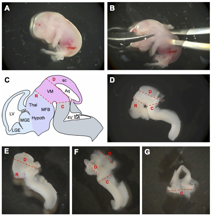
Molecular Diversity of Midbrain Development in Mouse, Human, and Stem Cells  - ScienceDirect
Molecular Diversity of Midbrain Development in Mouse, Human, and Stem Cells - ScienceDirect

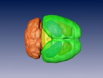
Is Athleticism Linked to Brain Size? - Neuroscience News

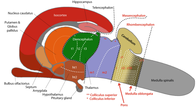
The Mouse Brainstem (Truncus encephali) | SpringerLink

Free Mouse coronal brain (midbrain) Icons, Symbols & Images | BioRender

GENSAT Project at Rockefeller University, Mouse Brain Atlas, Image Navigator

Primary Culture of Neurons Isolated from Embryonic Mouse Cerebellum |  Protocol (Translated to Italian)
Primary Culture of Neurons Isolated from Embryonic Mouse Cerebellum | Protocol (Translated to Italian)

Dissection of the ventral midbrain region from mice. Bregma coordinates...  | Download Scientific Diagram
Dissection of the ventral midbrain region from mice. Bregma coordinates... | Download Scientific Diagram
GENSAT Project at Rockefeller University, Mouse Brain Atlas, Image Navigator

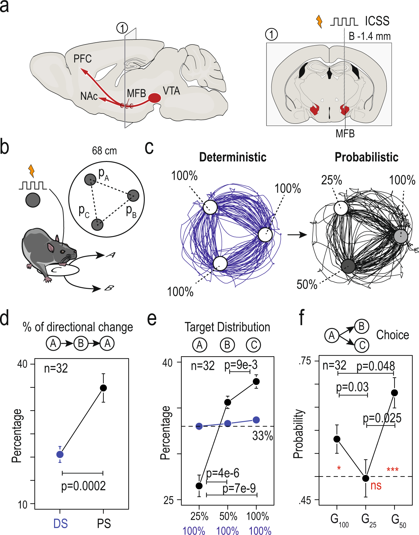
Chronic nicotine increases midbrain dopamine neuron activity and biases  individual strategies towards reduced exploration in mice | Nature  Communications
Chronic nicotine increases midbrain dopamine neuron activity and biases individual strategies towards reduced exploration in mice | Nature Communications

Morphology of mouse brain: olfactory bulbs, cerebral cortex, midbrain,  cerebellum, and spinal cord are labeled.
Morphology of mouse brain: olfactory bulbs, cerebral cortex, midbrain, cerebellum, and spinal cord are labeled.

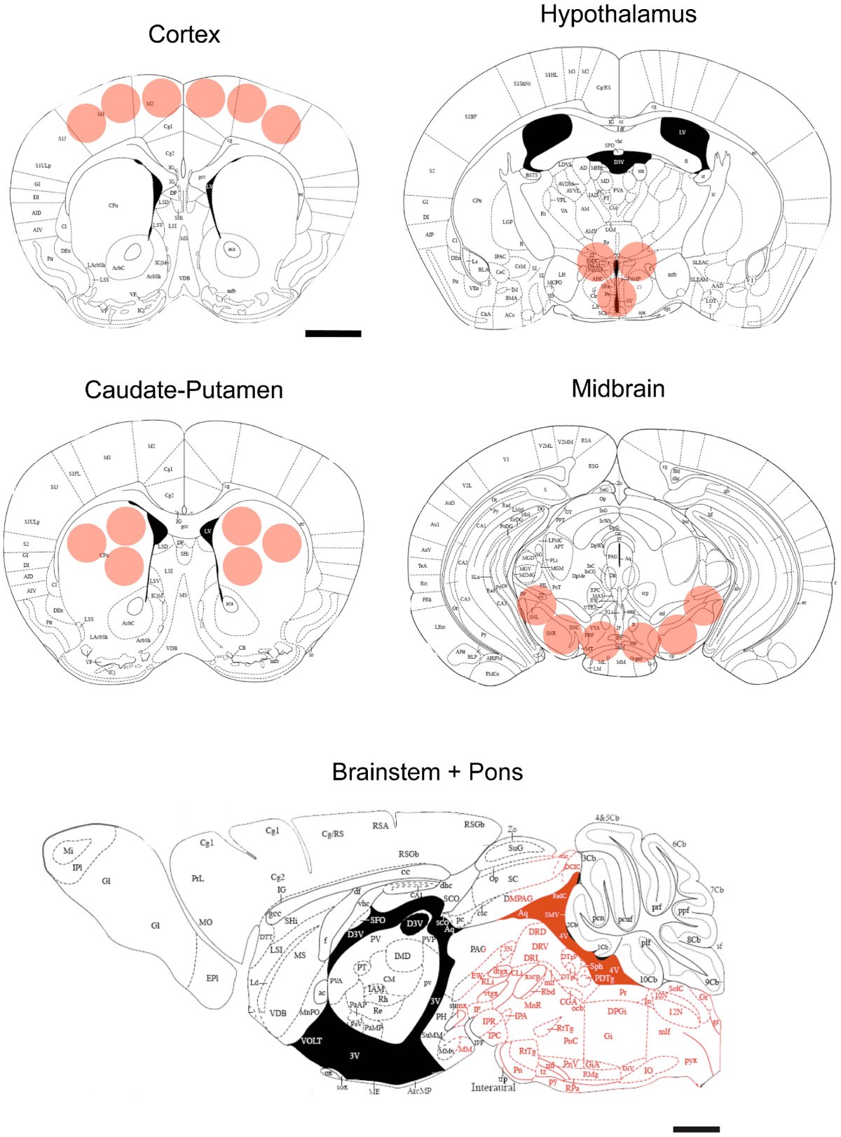
Normal Mouse Brain Proteome II: Analysis of Brain Regions by  High-resolution Mass Spectrometry | Cancer Genomics & Proteomics
Normal Mouse Brain Proteome II: Analysis of Brain Regions by High-resolution Mass Spectrometry | Cancer Genomics & Proteomics